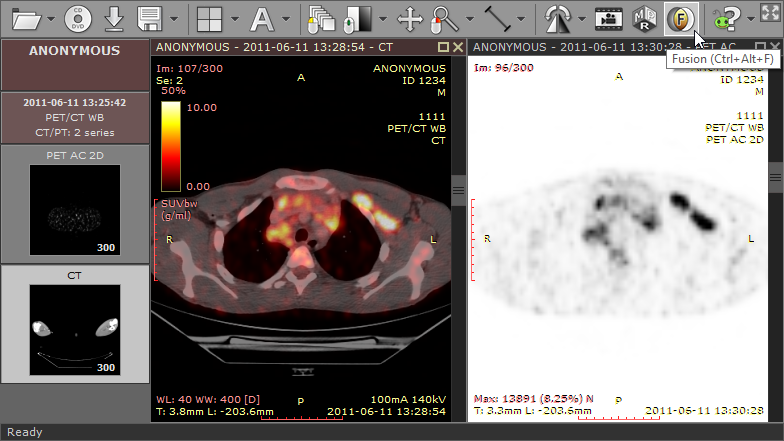
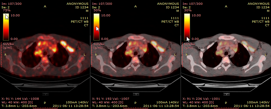
Image fusion allows you to overlay a color-mapped PET image onto a CT scan in order to obtain anatomical references for regions with increased FDG (fluorodeoxyglucose) uptake values.
This tool can also be applied to other imaging modalities, such as Magnetic Resonance, (e.g., DWI images can be fused with T1 or T2-weighted scans).
Enabling fusion
1) Use the screen splitting tool to open CT and PET series in two panels.
2) Activate the panel with CT series by clicking the image it contains or its title bar.
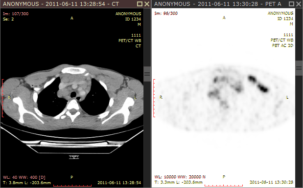
3) Click the Fusion button on the toolbar or use the Ctrl + Alt + F shortcut.
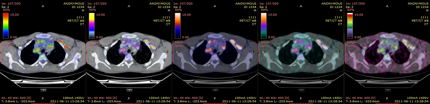
Color scale
The default color scale used for rendering a fused image is set to "Hot iron". It can be changed by clicking a bar with a color scale to the left side of the image.
The range of values represented on a color scale is adjusted by using the windowing tool in the panel with the series used as overlay (PET).
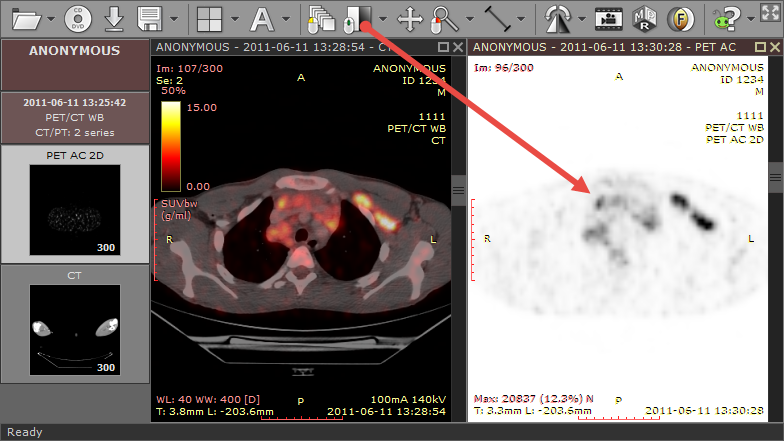
The opacity of fused images is adjusted by pressing the left mouse button over a color scale bar and dragging the mouse up to increase opacity or down to make the fused image more transparent.
SUV measurement
Use the ellipse tool in the panel with fused images to measure maximum, minimum and average values of SUVbw (Standardized Uptake Value calculated using body weight) for a specified area.
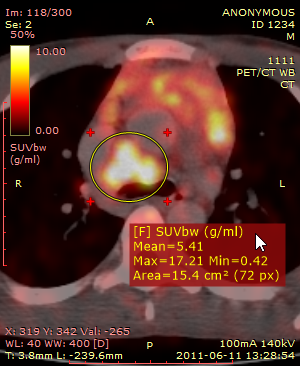
You can learn more about the SUV measurement in the 2D viewer section.