Online Store - RadiAnt DICOM Viewer - features and system requirements
| Basic Functionality |
| Desktop application for installation on PCs, laptops, and tablets running Windows systems |
| Windows 7/8/8.1/10/11 supported |
| Native ARM 64-bit version for the new generation of Windows on ARM devices (e.g., Surface Pro X) |
| No additional dependencies (.NET, Java, etc.) |
| Lightweight, compact application1 |
| Excellent performance with 32- and 64-bit versions that are both optimized for multi-core processors |
| Asynchronous reading (you can browse images as they are being opened) |
| Advanced memory management system that facilitates the concurrent opening of studies that contain thousands of images |
| Supported DICOM Formats |
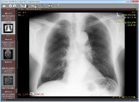
| Files from different imaging modalities: CR, DX, MG, CT, MR, PT, US, XA, NM, SC, SR |
| Monochromatic images (e.g., CR, CT, MR) |
| Color images (e.g., US, 3D reconstructions) |
| Static images (e.g., CR, MG, CT) |
| Dynamic sequences (e.g., XA, US) |
| Uncompressed images (little endian/big endian, implicit/explicit VR) |
| Compressed images (RLE, JPEG Lossy, JPEG Lossless, JPEG 2000, JPEG-LS) |
| Structured reports |
| Encapsulated PDF documents |
| MPEG4/MPEG2 DICOM videos |
| Access DICOM Studies |
| Open DICOM studies from CD/DVD/Blu-ray discs |
| Open DICOM studies from local and network folders |
| Open DICOM studies from USB drives |
| Open ZIP archives (unencrypted/encrypted) with DICOM files |
| Search and download DICOM studies (or selected series) from PACS locations (servers, workstations, modalities) |
| Accept and display studies pushed from other PACS locations |
| Local Archive |
| Store DICOM studies in a local database |
| Import DICOM studies from CD/DVD/Blu-ray discs |
| Import DICOM studies from local and network folders |
| Import DICOM studies from USB drives |
| Import ZIP archives (unencrypted/encrypted) with DICOM files |
| Import DICOM studies from PACS locations |
| Organize study collection using keywords |
| Multiple databases supported |
| Export list of studies to CSV file |
| Export Images |
| Export DICOM files to JPEG/BMP images |
| Export DICOM files to MP4/WMV movies |
| Export DICOM files in original format2 |
| Copy displayed image to Windows Clipboard |
| Send studies to PACS locations3 |
| Basic Tools |
| Perform fluid zooming |
| Perform fluid panning |
| Adjust brightness and contrast (window level/window width) |
| Negative mode |
| Apply window presets for computed tomography (lung, bone, etc.) |
| Apply precise window values (with SUVbw support for PET series) |
| Add your own window presets |
| Rotate (90 CW, 90 CCW, 180) |
| Flip (horizontal, vertical) |
| Apply image filters (sharpen, smooth, edge, emboss) |
| Display dynamic series/sequences (CINE) with option to adjust frames per second |
| Display DICOM overlays (annotations or graphic overlays included in the file) |
| Display DICOM file structure with searchable DICOM tags, their descriptions and values |
| Measurements/ROI |
| Measurement of segment length |
| Manual calibration of length measurements |
| Support for calibrated regions in ultrasound images |
| Measurement of mean, minimum and maximum parameter values (e.g. density in Hounsfield Units in computed tomography, SUVbw in PET) within circle/ellipse and its area |
| Measurement of area and perimeter of a closed polygon |
| Measurement of open polygon length |
| Measurement of angle value |
| Measurement of Cobb angle value |
| Measurement of deviation distance |
| Arrow tool for annotations |
| Pencil tool for freehand drawing |
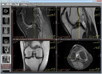
| Compare Series |
| Compare multiple series in the same or different windows |
| Automatic synchronization between series with images acquired in the same plane (e.g., computed tomography series before and after contrast media administration)4 |
| Manual synchronization between series from different studies that have a similar patient orientation |
| Cross-reference lines in series with different image planes (e.g., magnetic resonance study)4 |
| 3D cursor tool |
| Split multi-sequence series into separate panels |
| Advanced Tools |
| 2D MPR (orthogonal multiplanar reconstructions) |
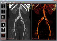
| Fusion of series with different modalities (e.g., PET-CT) or with different protocols (e.g., MR T1/T2 – DWI) |
| Time-intensity curves (TIC, e.g., for breast MRI) |
| 3D MPR (oblique multiplanar reconstruction) with MIP (maximum intensity projection), MinIP (minimum intensity projection) and Avg (average) modes |
| 3D VR (volume rendering) |
| 3D snapshots for quick saving and restoring 3D VR views |
| Creation of quick movies (simple rotations) and advanced 3D snapshot-based videos with volume-rendered objects |
| Export 3D models to STL files |
| GPU acceleration for 3D VR and 3D MPR/MIP5 |
| DSA mode (digital subtraction angiography) with auto and manual pixel-shift, split mask and magic mask |
| Interface |
| A simple and intuitive interface with full-screen and distraction-free modes |
| Multi-touch support for Windows 8/8.1/10/11 touch-enabled devices |
| Multilingual interface—more than 30 translations available6 |
| Customizable keyboard shortcuts |
| Integration with third-party systems via command-line arguments and the radiant:// URL protocol |
| Minimum system requirements |
| Microsoft Windows system (Windows 7, Windows 8, Windows 8.1, Windows 10 and Windows 11 are supported) |
| Intel or AMD 1GHz or faster processor |
| 1GB of RAM |
| 10MB of available hard-disk space for installation; additional free space required for the local database of studies and image caching |
| 1024 x 768 screen resolution |
| Recommended system requirements |
| Microsoft Windows 10 or Windows 11 system |
| Intel or AMD 3GHz or faster processor with four or more cores |
| 4GB of RAM (8GB for 3D viewing) or more |
| Fast SSD system drive |
| 1920 x 1080 (or higher) screen resolution |
| NVIDIA graphics card (10 series or newer) for GPU-accelerated 3D VR and MPR |
1 The installer size of RadiAnt DICOM Viewer is 7MB; the disk space occupied after installation is 10MB.
2 It is not possible to export secondary images obtained using Multiplanar Reconstructions (MPR), 3D Volume Rendering, Fusion, or Time-intensity Curve (TIC) tools to DICOM files.
3 Available with the DCMTK add-on package installed.
4 The series have to belong to the same study.
5 Only available with the 64-bit version of RadiAnt DICOM Viewer. Supported NVIDIA graphics card required.
6 RadiAnt DICOM Viewer and RadiAnt DICOM Viewer CD/DVD are distributed with English and Polish language support. Other translations (Arabic, Brazilian Portuguese, Bulgarian, Chinese, Chinese [Taiwan], Croatian, Czech, Danish, Dutch/Flemish, French, German, Greek, Hungarian, Indonesian, Italian, Japanese, Korean, Mongolian, Norwegian, Persian, Portuguese, Romanian, Russian, Serbian, Slovak, Spanish, Swedish, Turkish, Ukrainian) have been created by the community and are available for download from https://www.radiantviewer.com/translations/. Not all translations have been updated for the newest version of the viewer.
RadiAnt DICOM Viewer is not certified as a medical product (there are no FDA/CE-1 or any other certifications for the software).